Research

DNA Repair

Laser Scissors and Tweezers

A gif of a sciam

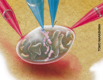
Combinations of laser scissors and tweezers can make it possible to perform subtle subcellular manipulations.  In a procedure that should be feasible within a decade, two tweezers beams (pink) hold a cell firmly in place.  One scissor beam (lighter blue) penetrates the cell to delete a faulty gene (red).  A second scissors beam (dark blue) cuts a hole in the cell membrane through which a competent genetic sequence (black dots) can pass.  Clones of the genetically altered cell could be then produced and transplanted into the body for therapeutic use.  (Berns, MW., “Laser Scissors and Tweezers,” Scientific American, Apr 1998)

Neural Photonics

Laser Scissors and Tweezers

Image of microtuble

One of the most exciting developments in cell and developmental biology over the past has been the creation of genetic fluorescent fusion proteins, as exemplified by green fluorescent protein (GFP). This has permitted visualization of structures in live cells not previously possible. A focus of our present and future research is to use fluorescent fusion proteins to visualize and study cell structures that can be manipulated with laser scissors and tweezers. We are already applying this combined systems approach to problems of cellular motility, particularly with respect to organelles of the mitotic spindle (kinetochores, centrosomes, microtubules; Botvinick et al, Biophysical Journal, Dec 2004). This approach will be used to study other organelles such as nucleoli, golgi, and the cell membrane.

Cell Motility

Laser Tweezers: Cellular Motility and Biomechanics

Image of Optical Trapping

Laser scissors are used to alter subcellular organelles involved in cell motility (microtubules, chromosome centromeres, centrosomes/centrioles). Laser tweezers are used to apply specific forces to structures of the mitotic spindle and cell cytoskeleton in order to study dynamic interactions and biomechanics in live cells. Fluorescence recovery after photobleaching (FRAP) is being used to examine the functional biochemistry of these structures. We are studying the mobility and turnover of specific chemical species (MAD1 and MAD2) at the cell kinetochore by FRAP (Shah et al, Cur. Biol., Jun 2004). We are continuing to study these molecules using fluorescence correlation spectroscopy (FCS), which permits the measurement of the change in molecular concentration at different regions of the cell during the cell cycle (Wang et al, J. Biomed. Optics., Mar-Apr 2004).

Instrumentation

Robolase: Internet-Based Cooperative Microscopy

A new focus of our program has been to engineer, design, and build a fully automated robotic laser microscope (Robolase) that can be operated in near real-time from remote sites via the internet. This will be both a research and an instructional tool. The prototype Robolase I was developed as a multitask system that employed both laser scissors (microablation) and laser tweezers (trapping). The system can be operated via the internet with control transferred to the user via a web page that presents a control panel allowing: (1) laser selection (tweezers or scissors) and operation, (2) selection of microscope imaging system (bright field, phase contrast, fluorescence), and (3) full control of the microscope in the x, y, and z planes as well as objective rotation and filter combination selection. This system Robolase II has been installed and is operational between the UCI and UCSD bioengineering programs. Robolase II and its future iterations will present even greater selection and control features for the user or collaborator. Robolase III is underconstruction and will allow us to study and manipulate cells and tissues like never before.

Mechanisms of Photon Interaction

In order to effectively utilize the above approach, it is necessary to understand the mechanisms of photon interaction, as they may vary greatly depending up laser fluence, wavelength, and absorption.  With the availability of nanosecond, picosecond and femtosecond lasers, we have undertaken studies to delineate the mechanisms of photon interaction so that they may be used to produce desired photo-physical and biological effects.  (See discussion Botvinick et al, Biophysical Journal, Dec 2004).

A diagram of photon interaction

Ray diagram of a bead trapped in an optical trap. A) The focus of the laser is above the center of the bead, creating an upward gradient force, balanced by a downward scattering force (not shown). This results in a stable laser trap. B) The focus is below the center creating a downward gradient force and in conjunction with a downward scattering force will cause the bead out of the trap. C) The focus is to the left of the center, creating a gradient force to the left. (Ashkin, 1998)

For example, it has been possible to employ two photon absorption by an ethidium dye to selectively inactivate ribosomal genes by two photon photochemistry (Berns et al, Proc. Natl. Acad. Sci., Aug 2000).   In addition other fluorescent dyes are being developed that can be optically switched on and off via conversion from a polar to non-polar state.  These dyes may serve as intracellular and extracellular biosensors (Berns et al., Photochemistry and Photobiology B, in press).

Using laser tweezers we are studying intracellular signal transduction by applying a laser trapping force to micron-size beads that are attached to the cell surface by selective ligand binding.  The stretching force applied to the cell surface results in a cascade of intracellular events involving the activation of the sarc protein.  A time-based gradient of enzyme activation can be detected at points in the cell distal to the site of the applied force (Wang et al, Nature, Apr 2005).   This approach is being used to study mechanotransduction in cells, and should be applicable to other signal transduction pathways. (Movie 1) (Movie 2)

A photo of a robolase system setup

Robolase I system setup

Experiments will be able to be conducted from any of the UC campuses and from any location around the world with internet access. With image compression and feature extraction techniques, near real-time operation will soon be possible. Collaborative projects are already underway between UCSD, UCI, and UC Berkeley, and planning discussions are underway with bioengineering programs at UC Riverside, UC Davis and the University of Massachusetts. The initial goal will be to establish both educational and research links with the different bioengineering programs within the UC system as part of the Bioengineering MRU. The idea is to make sophisticated, expensive, cutting edge technology available for training of students, as well as for research projects of faculty who may not have access to the technology.

A gif of scientists reading red blood cells

Collaborating researchers at the University of Queensland, Australia (UQ) were able to ablate red blood cells through the internet using our Robolase II setup at the University of California, San Diego (UCSD)